Eye anatomy
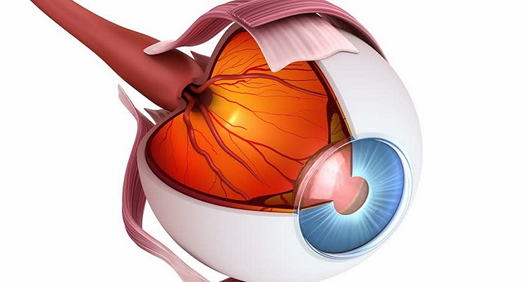
The eye is one of the most complex organs in the body. Responsible for taking light signals to the brain, it functions as a natural camera that is an extension of the brain. Eyeball is made up of 3 main layers. Outermost layer is called Sclera, middle layer is called Uvea.It has several layers that are equivalent to the lens of the camera, the film, and the lens cover. Remember that any part of the eye can be involved in the disease process, and may require a subspecialist for respective involved part of eye.
Cornea
The cornea is the clear protective coating on the front of the eye, like the glass on a watch, that allows light to pass through it without distortion. It covers the colored iris. The lens of the eye focuses the images transmitted through the cornea to the retina. Therefore, it must be clear and regularly shaped to give good vision. These images are then transferred via the optic nerve to the brain, where sight is interpreted. A "scratched" cornea is extremely painful and it seeks urgent treatment.